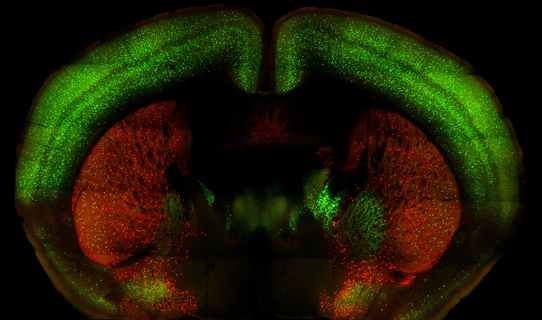
The mouse cortex and striatum. PV interneurons are stained green and cholinergic interneurons are stained red.
|
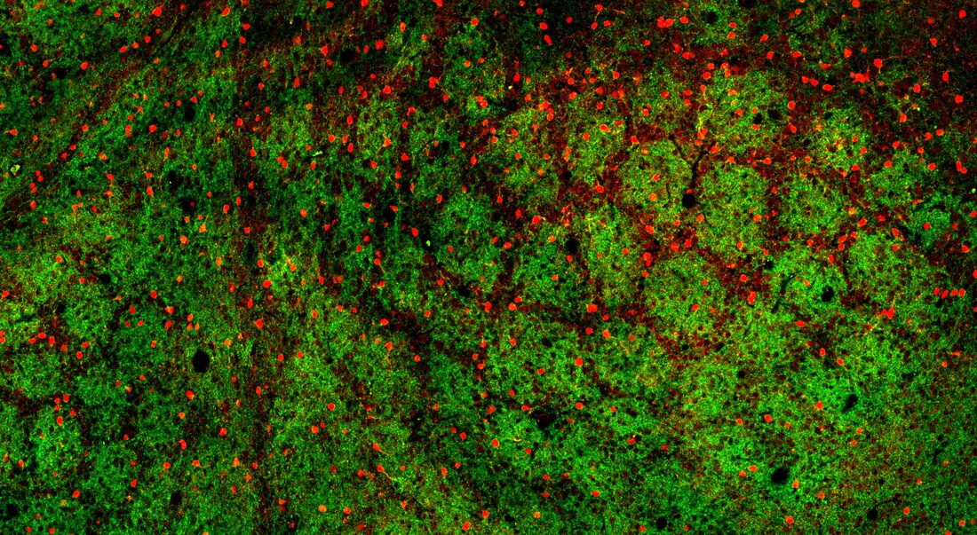
vGlut2 positive axons terminating in the mouse barrel cortex. PV interneurons are stained in red.
|
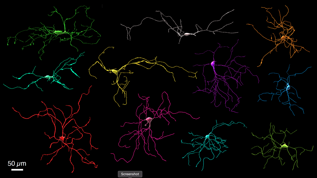
Neurobiotin dye fills detailing different populations of cholinergic interneurons in the striatum
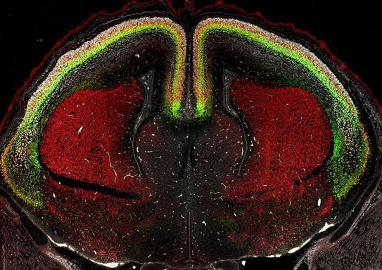
Coronal section through the mouse embryonic cortex and striatum. TBR1 (green) marks cortical layer 6, CTIP2 in red marks cortical layer 5, and Satb2 in white marks layers 2-4.
|
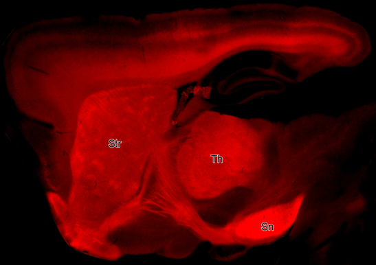
Brain section from a Drd1-Cre:Ai14 mouse detailing striatal (str) projections to the substantia nigra (Sn). Th=thalamus
|
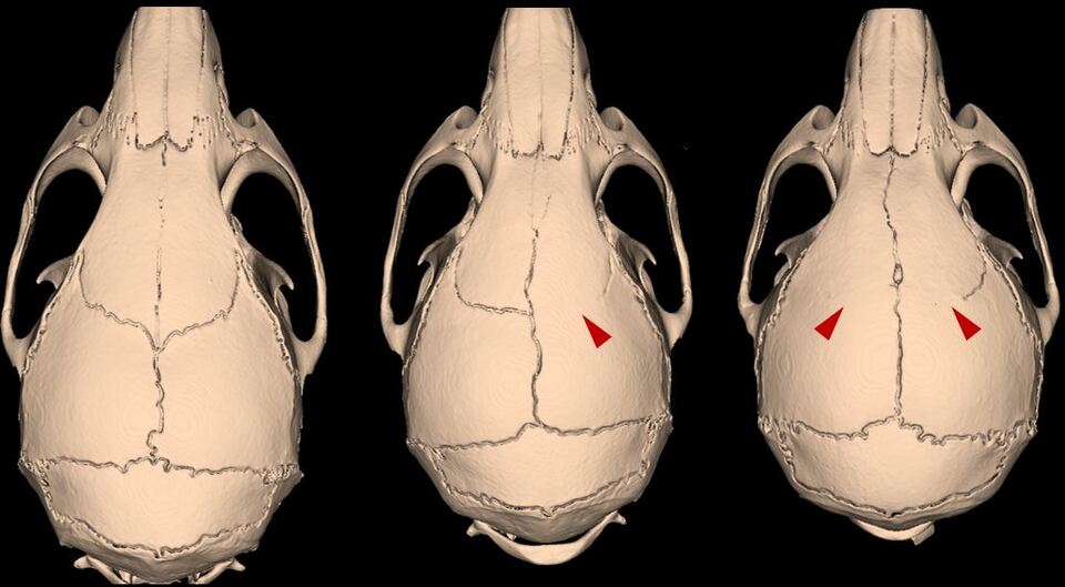
Computed Tomography scans of a normal mouse (left) and affected littermates who have craniosynostosis due to heterozygous loss of Twist1 in the cranial sutures. Red arrows denote coronal suture fusion, which can be present unilaterally or bilaterally.
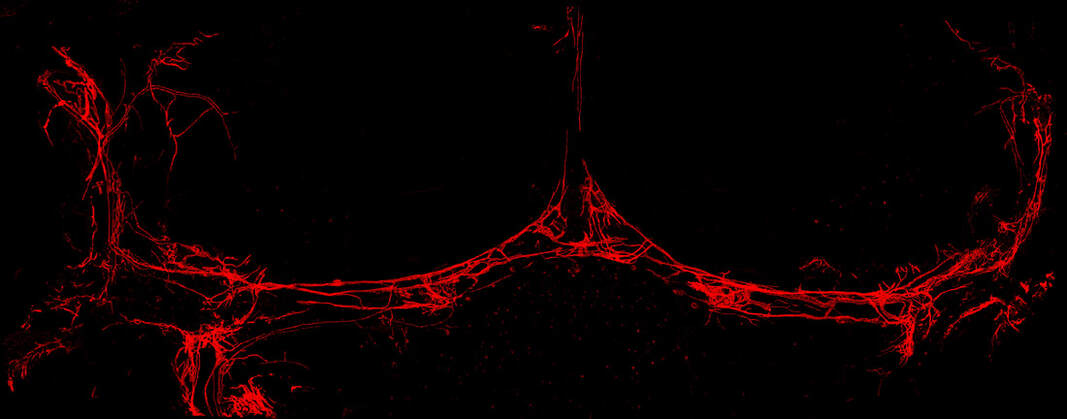
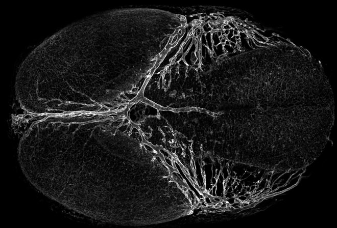
A dorsal view of meningeal lymphatic networks in dura as visualized using Prox1-tdTomato transgenic mice.
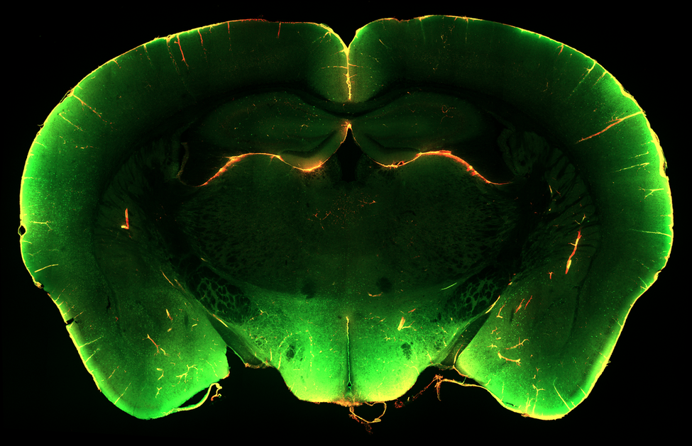
Coronal section through an adult mouse brain. FITC-conjugated dextran is seen entering the brain through perivascular spaces following injection via the cisterna magna. This process is essential for waste clearance in the CNS.
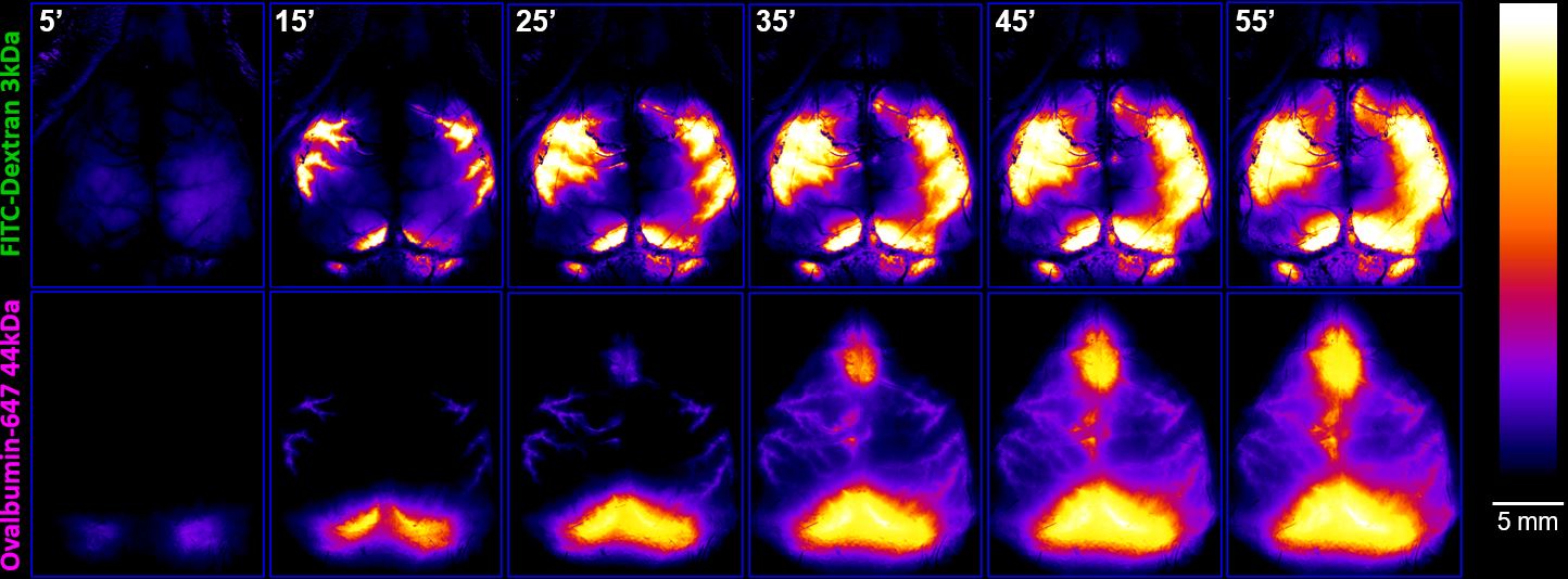
Time lapse imaging of cerebrospinal fluid circulation in an adult mouse using live transcranial imaging. Two different sized molecular tracers were injected into the cerebral spinal fluid. Tracking the tracers over time allows us to measure the velocity.
Top-down dorsal view of an embryonic day 12.5 mouse brain, with the primitive venous sinuses and venous capillaries labeled with endomucin. The mature venous sinuses arise from skull-induced venous growth and remodeling at this stage.
Three-dimensional x-ray microscopy showing an adult mouse skull. (credit: Pat Buckendahl, RUMIC, RTS)
Loss of balanced retinoic acid signaling from the meninges causes cortical folding (on left) in mice
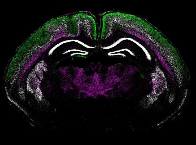
Cortical layers: Satb2 (green), Ctip2 (white), FoxP2 (magenta)
Cortical layers: Satb2 (green), Ctip2 (white), FoxP2 (magenta)
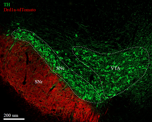
Section through the midbrain of an adult mouse showing axons from striatal medium spiny neurons (red) innervating the Substantia Nigra Reticulata (SNr). Green labels tyrosine hydroxylase (TH) expressing dopaminergic neurons and their projections in the Substantia Nigra Compacta (SNc) and Ventral Tegmental Area (VTA)